
Researchers have unveiled a groundbreaking software tool that allows for unprecedented insight into the 3D dynamics of biological structures. This interactive, dynamic tool provides researchers with advanced cutaway views of 3D images, making it possible to analyze the never-before-seen processes of embryonic heart development using optical coherence tomography (OCT) images.
Led by Shang Wang from Stevens Institute of Technology, the team’s work is set to significantly impact both the study of congenital heart diseases—one of the most common birth defects—and strategies for regenerating heart tissue after a heart attack. Their research, published in Biomedical Optics Express by the Optica Publishing Group, introduces the “clipping spline,” a new open-source software that offers an intuitive way to visualize complex 3D structures.
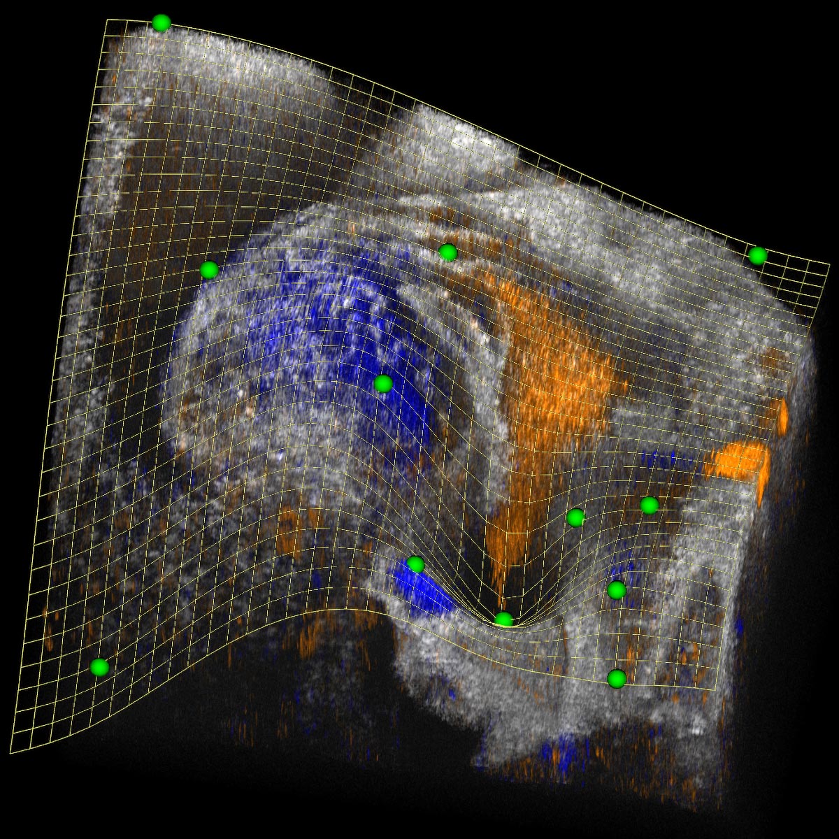
The clipping spline software was developed to address a critical need in biomedical imaging—specifically, the ability to visualize complex, 3D structures in a single cutaway view. Traditionally, volume clipping—removing parts of a 3D image to reveal its internal structure—has been limited by the geometry of cutting tools, which often fail to produce intricate views of biological structures. The clipping spline overcomes this by using a smooth, adjustable surface known as a “thin plate spline” (TPS), a mathematical model that can dynamically cut through 3D volumes with high precision.
“3D imaging is already essential in biomedicine, but when we consider the temporal aspect—turning 3D images into 4D—the possibilities become even greater,” said Andre Faubert, a research associate on the team. While the clipping spline was first demonstrated with 4D OCT data, it can be used for volumetric images from any imaging modality, with applications in both biological research and clinical medicine.
The researchers initially applied the clipping spline to study the development of the embryonic mouse heart, focusing on the critical “cardiac looping” stage. During this stage, the heart tube bends and twists, forming complex shapes and generating specific blood flow patterns. However, understanding the dynamics of this process has been challenging due to the complexity of the heart’s development and a lack of suitable visualization tools.
“Heart development, especially during the looping stage, is crucial because defects in this process are responsible for many congenital heart defects,” Wang explained. “But until now, there were very few tools to visualize and analyze these complex changes as they happen.”
By using the clipping spline, the researchers were able to track the heart’s development over 12.8 hours of embryonic growth, capturing 712 time points. This provided a comprehensive, real-time view of the heart’s biomechanics, allowing them to study how blood flow patterns are generated and how different regions of the heart merge to form key structures such as the sinus venosus, which directs blood into the developing heart.
The clipping spline tool’s ability to provide cutaway views of the complex embryonic heart allowed the researchers to observe multiple sections of the heart simultaneously. This offered a larger and more integrated perspective of its development compared to previous methods. They were able to gain insights into the merging of the inflow tracts and how the shape and movement of the heart tube influence blood flow dynamics.
“This is truly an amazing achievement,” said Wang. “By visualizing these developmental processes in greater detail, we can generate new hypotheses and insights into how the mammalian heart develops. This research is not only essential for understanding congenital heart diseases but also has broader implications for fields like cancer research and regenerative medicine.”
The researchers believe the clipping spline software has the potential to transform the way scientists and clinicians approach 3D imaging and analysis. By simplifying and streamlining complex volume clipping, this tool can be widely applied across various imaging modalities in both research and clinical settings.
The software is now ready for broader use within the biomedical imaging community, with further development underway to enhance its image-processing capabilities. The team’s next step is to apply the clipping spline to explore additional aspects of heart development, including the role of biomechanics and tissue interactions in organ formation.
“Understanding how biological structures evolve is fundamental to advancing medicine, and the clipping spline opens up new possibilities for exploring the dynamics of this development,” said Faubert. “We are excited to see how it will be applied in a variety of biomedical contexts in the future.”
By Impact Lab