
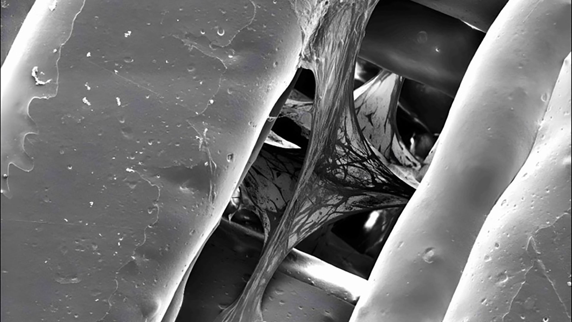
In a groundbreaking development, researchers have harnessed the power of low-cost FDM 3D printing technology to create scaffolds that closely mimic the mineral properties of natural bone tissue. By combining poly(lactic-co-glycolic acid) (PLGA) with hydroxyapatite (HA), they’ve created a base structure that offers a realistic environment for bone research. This innovative method significantly lowers the barrier to entry for labs, as it eliminates the need for expensive bioprinters traditionally required for such work.
While high-end bioprinters can cost hundreds of thousands of dollars, this new technique shows that even inexpensive desktop 3D printers can be used to produce functional scaffolds that are suitable for complex biomedical research. This democratization of technology could allow a wider range of labs, especially those with limited funding, to engage in advanced bone tissue research.
The true innovation lies in the combination of these 3D-printed scaffolds with bone marrow mesenchymal stem cells (BM-MSCs)—cells that are capable of differentiating into various types of tissue. When applied to the scaffolds, these stem cells are able to move within the structure’s microscopic pores, absorbing nutrients and organizing themselves in a way that closely resembles the natural processes of bone formation.
The 3D-printed structure, with its highly controlled porosity, provides an ideal environment for these cells to thrive and potentially develop into bone-like tissue. This has profound implications for both cancer research and drug development, as the method could create more accurate in-vitro models for studying bone diseases and testing new treatments.
Beyond advancing cancer research, this new 3D printing method has the potential to drastically reduce the need for animal testing, a practice that is both costly and ethically controversial. By providing a more realistic in-vitro model for studying bone tissue and its diseases, researchers can more efficiently develop and test treatments without relying on animal subjects.
This technology also opens the door to broader applications in bone-related diseases, such as osteoporosis and osteoarthritis, by providing researchers with more effective ways to study and treat conditions that affect bone tissue. With the ability to easily reproduce scaffolds with the same material properties, the technique could help in the development of personalized medicine and targeted therapies for individuals suffering from these ailments.
This breakthrough in 3D-printed bone scaffolds represents a significant leap forward in biomedical research. It combines the affordability and accessibility of desktop 3D printing with the complexity needed to replicate the natural structure of bone tissue. As the technology continues to evolve, it could pave the way for a new era in tissue engineering and regenerative medicine, making it easier and more cost-effective to develop treatments for a variety of bone diseases and advancing the study of cancer therapies.
By Impact Lab