“Braingeneers” automate growth of brain tissue organoids on a chip

NOVEL SYSTEM CAN INCREASE REPRODUCIBILITY IN CEREBRAL ORGANOID RESEARCH AND SHOWS PROMISE FOR LOWERING LEVELS OF CELLULAR STRESS.

By  Eleanor Garth

A team of engineers at UC Santa Cruz has developed a new method for remote automation of the growth of cerebral organoids – miniature, three-dimensional models of brain tissue grown from stem cells.

Longevity.Technology: Cerebral organoids allow researchers to study and engineer key functions of the human brain with a level of accuracy not possible with other models. This has implications for understanding brain development and the effects of pharmaceutical drugs for treating neurodegenerative diseases or other diseases of aging.

Research on aging has primarily been conducted using cell cultures, yeast, C elegans and animal models (flies, mice). However, longevity research is committed to unpicking the pathways that regulate aging and developing interventions that could slow biological aging and delay the onset and progression of age-related diseases in humans. So, while we have a wealth of non-human data, how much if it is directly applicable to extending human healthspan?

Organoids enable interventional studies that are difficult or impossible to conduct in humans, while at the same time providing valuable human data – their potential as a significant preclinical model tool is enormous.

In this new study, which has been published in the journal Nature Scientific Reports, researchers from the UCSC Braingeneers group (probably the best name you’ll read this week!) detail their automated, internet-connected microfluidics system, called “Autoculture”; this system precisely delivers feeding liquid to individual cerebral organoids in order to optimise their growth without the need for human interference with the tissue culture.

Continue reading… ““Braingeneers” automate growth of brain tissue organoids on a chip”

Scientists Have Established a Key Biological Difference Between Psychopaths and Normal People

The research found that the striatum region of the brain was on average ten percent larger in psychopathic individuals compared to a control group of individuals that had low or no psychopathic traits.

A new study has shown that psychopathic people have a bigger striatum area in their brain.

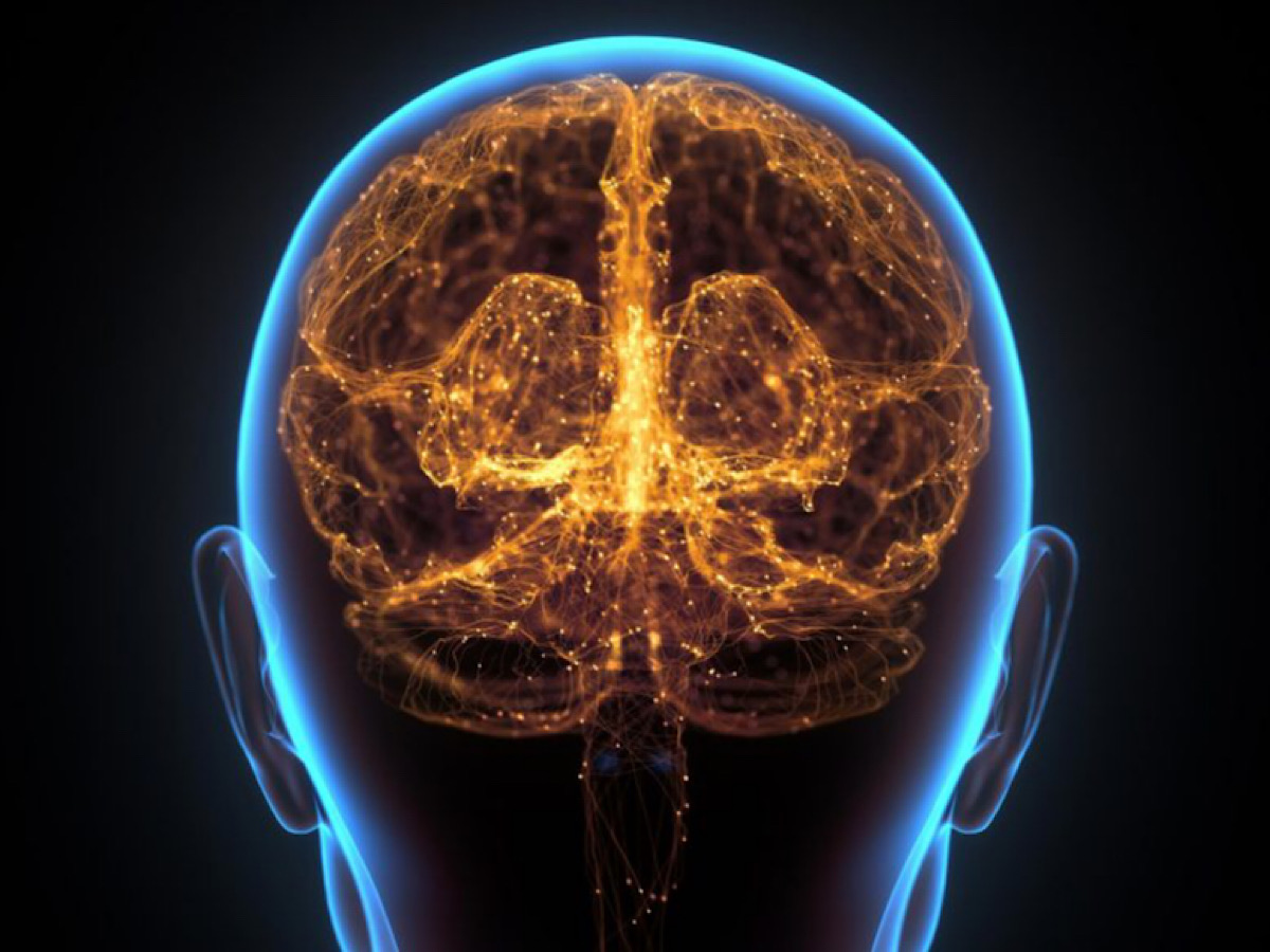
Neuroscientists using MRI scans discovered that psychopathic people have a 10% larger striatum, a cluster of neurons in the subcortical basal ganglia of the forebrain, than regular people. This represents a clear biological distinction between psychopaths and non-psychopathic people.

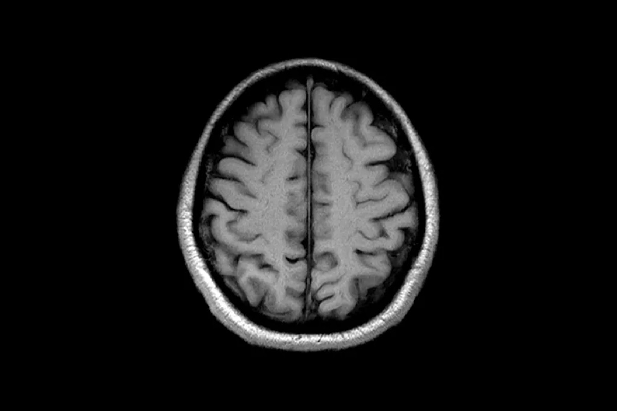
Neuroscientists from Nanyang Technological University (NTU Singapore), the University of Pennsylvania, and California State University have discovered a biological distinction between psychopaths and non-psychopaths. Using magnetic resonance imaging (MRI) scans, scientists discovered that the striatum, an area of the forebrain, was 10% bigger in psychopathic people compared to a control group of individuals with low or no psychopathic traits.

Psychopaths, or those with psychopathic qualities, are people who have an egotistical and antisocial disposition. This is often characterized by a lack of guilt for their actions, a lack of empathy for others, and, in some cases, criminal tendencies.

The striatum, which is part of the forebrain, the subcortical region of the brain that encompasses the whole cerebrum, coordinates numerous elements of cognition, including motor and action planning, decision-making, motivation, reinforcement, and reward perception.

Previous research has shown that psychopaths have overactive striatum, but the influence of its size on behavior has yet to be confirmed. The new research demonstrates a significant biological difference between people who exhibit psychopathic tendencies and those who do not. While not all people with psychopathic qualities end up violating the law, and not all criminals satisfy the criteria for psychopathy, there is a strong association. There is also significant evidence that psychopathy is associated with more aggressive behavior.

Continue reading… “Scientists Have Established a Key Biological Difference Between Psychopaths and Normal People”

Google has mapped a piece of human brain in the most detail ever

Around 4000 nerve fibres connect to this single neuronGoogle/Lichtman Laboratory

By  Michael Marshall

Google has helped create the most detailed map yet of the connections within the human brain. It reveals a staggering amount of detail, including patterns of connections between neurons, as well as what may be a new kind of neuron.

The brain map, which is freely available online, includes 50,000 cells, all rendered in three dimensions. They are joined together by hundreds of millions of spidery tendrils, forming 130 million connections called synapses. The data set measures 1.4 petabytes, roughly 700 times the storage capacity of an average modern computer.

The data set is so large that the researchers haven’t studied it in detail, says Viren Jain at Google Research in Mountain View, California. He compares it to the human genome, which is still being explored 20 years after the first drafts were published.

It is the first time we have seen the real structure of such a large piece of the human brain, says Catherine Dulac at Harvard University, who wasn’t involved in the work. “There’s something just a little emotional about it.”

Continue reading… “Google has mapped a piece of human brain in the most detail ever”

If You Transplant a Human Head, Does Its Consciousness Follow?

MAX G. LEVYSCIE

In her new book, Brandy Schillace recalls the unbelievable legacy of a Cold War era neurosurgeon’s mission to preserve the soul.

BRANDY SCHILLACE SOMETIMES writes fiction, but her new book is not that. Schillace, a medical historian, promises that her Cold War-era tale of a surgeon, neuroscientist, and father of 10 obsessed with transplanting heads is true from start to finish. 

Schillace came across the story behind her book, Mr. Humble and Dr. Butcher, somewhat serendipitously: One day, her friend, Cleveland neurologist Michael DeGeorgia, called her to his office. He quietly slid a battered shoebox toward her, inviting her to open it. Schillace obliged, half-worried it might contain a brain. She pulled out a notebook—perhaps from the ‘50s or ‘60s, she says—and started to leaf through it.

“There’s all these strange little notes and stuff about mice and brains and brain slices, and these little flecks,” Schillace says. “I was like, ‘What … what are all these marks?’”

Continue reading… “If You Transplant a Human Head, Does Its Consciousness Follow?”

Ultrasound Imaging Technique Allows Scientists To Read Minds

The team successfully tested the method on non-human primates and read out their brains’ intentions.

By  Fabienne Lang 

A new type of brain-machine interface (BMI) that’s minimally invasive can read out the brain’s intentions using ultrasound technology. 

A collaborative team of researchers at Caltech developed the system that can read brain activity corresponding to the planning of movement. 

The team’s study was published in the journal Neuron on Monday 22 March.

Neuroscientists working on BMIs in order to map out the brain’s activity to corresponding movements will be having a field day thanks to this new study. Typically, these devices read and interpret brain activity and link it up to a computer or machine. 

However, these devices typically require invasive brain surgery, which many patients aren’t willing to partake in. 

The news of this new technology, which uses functional ultrasound (fUS) technology accurately maps out neural activity from its source deep within the brain at a resolution of 100 micrometers. 

Continue reading… “Ultrasound Imaging Technique Allows Scientists To Read Minds”

New technology ‘retrains’ cells to repair damaged brain tissue in mice after stroke

Graduate research associate Jordan Moore reviews brain MRI images of mice in the nanomedicine lab at The Ohio State University College of Medicine. In a new study, researchers demonstrate the potential of a new cell therapy to reverse damage from ischemic stroke by regrowing blood vessels and healthy brain tissue. Credit: The Ohio State University Wexner Medical Center

by The Ohio State University

Most stroke victims don’t receive treatment fast enough to prevent brain damage. Scientists at The Ohio State University Wexner Medical Center, College of Engineering and College of Medicine have developed technology to “retrain” cells to help repair damaged brain tissue. It’s an advancement that may someday help patients regain speech, cognition and motor function, even when administered days after an ischemic stroke.

Engineering and medical researchers use a process created by Ohio State called tissue nanotransfection (TNT) to introduce genetic material into cells. This allows them to reprogram skin cells to become something different—in this case vascular cells—to help fix damaged brain tissue.

Study findings published online today in the journal Science Advances.

In this mouse study, cells were ‘pre-conditioned’ with specific genes and injected into the stroke-affected brains, where they promoted the formation of new blood vessels via reprogramming and the repair of damaged brain tissue.

“We can rewrite the genetic code of skin cells so that they can become blood vessel cells,” said Daniel Gallego-Perez, an assistant professor of biomedical engineering and surgery at Ohio State who is leading the research. “When they’re deployed into the brain, they’re able to grow new, healthy vascular tissue to restore normal blood supply and aid in the repair of damaged brain tissue.”

Researchers studied the process in mice and found that those treated with this innovative cell therapy regained 90% of their motor function. MRI scans showed damaged areas of the brain were repaired within a few weeks.

Continue reading… “New technology ‘retrains’ cells to repair damaged brain tissue in mice after stroke”

GUT MICROBES COULD BE THE FUTURE OF BRAIN HEALTH

“We might end up being able to correct a behavioral deficiency by what we put in a milkshake.”

By KATIE MACBRIDE

THE EVIDENCE FOR A CONNECTION between gut health and brain health is becoming increasingly hard to ignore. A new study adds to the mountain: In the paper, a team of scientists at Baylor College of Medicine link gut bacteria to specific brain conditions. 

But beyond this, the team may have unlocked how to leverage the connection to treat brain conditions that affect social behavior. 

WHAT’S NEW — The new research suggests hacking that connection via the vagus nerve by changing the composition of microbes in the gut. This nerve functions as a kind of fiber-optic cable that carries messages between the gut and the brain. Specifically, the team behind this paper looked at the microbiome’s role in hyperactivity seen in mice lacking a gene associated with autism. What they found suggests that altering the population of gut microbiota through food may in turn alter behavior. 

Continue reading… “GUT MICROBES COULD BE THE FUTURE OF BRAIN HEALTH”

What can AI learn from Human intelligence?

463B4E56-6F28-4B0E-8618-9FB011B66E7F

At HAI’s fall conference, scholars discussed novel ways AI can learn from human intelligence – and vice versa.

Can we teach robots to generalize their learning? How can algorithms become more commonsensical? Can a child’s learning style influence AI?

Stanford Institute for Human-Centered Artificial Intelligence’s fall conference considered those and other questions to understand how to mutually improve and better understand artificial and human intelligence. The event featured the theme of “triangulating intelligence” among the fields of AI, neuroscience, and psychology to develop research and applications for large-scale impact.

HAI faculty associate directors Christopher Manning, a Stanford professor of machine learning, linguistics, and computer science, and Surya Ganguli, a Stanford associate professor of neurobiology, served as hosts and panel moderators for the conference, which was co-sponsored by Stanford’s Wu-Tsai Neurosciences Institute, Department of Psychology, and Symbolic Systems program.

Speakers described cutting-edge approaches—some established, some new—to create a two-way flow of insights between research on human and machine-based intelligence, for powerful application. Here are some of their key takeaways.

Continue reading… “What can AI learn from Human intelligence?”

The most fundamental skill: Intentional learning and the career advantage

2189CC1D-957F-418F-8714-FB7334EF7A82

Learning itself is a skill. Unlocking the mindsets and skills to develop it can boost personal and professional lives and deliver a competitive edge.

The call for individuals and organizations alike to invest in learning and development has never been more insistent. The World Economic Forum recently declared a reskilling emergency as the world faces more than one billion jobs transformed by technology. Even before COVID-19 emerged, the world of stable lifetime employment had faded in the rearview mirror, replaced by the expectation that both executives and employees must continually refresh their skills. The pandemic has only heightened the urgency of doubling down on skill building, either to keep up with the speed of transformation now underway or to manage the particulars of working in new ways.

Despite this context—and the nearly constant refrain for people to adapt to it by becoming lifelong learners—many companies struggle to meet their reskilling goals, and many individuals struggle to learn new and unfamiliar topics effectively. We believe that an underlying cause is the fact that so few adults have been trained in the core skills and mindsets of effective learners. Learning itself is a skill, and developing it is a critical driver of long-term career success. People who have mastered the mindsets and skills of effective learning can grow faster than their peers and gain more of the benefits from all the learning opportunities that come their way.

This article, supported by research and our decades of experience working as talent and learning professionals, explores the core mindsets and skills of effective learners. People who master these mindsets and skills become what we call intentional learners: possessors of what we believe might be the most fundamental skill for professionals to cultivate in the coming decades. In the process they will unlock tremendous value both for themselves and for those they manage in the organizations where they work.

Continue reading… “The most fundamental skill: Intentional learning and the career advantage”

The surprising upsides of worrying

 88EC81AB-A29D-471D-AFAB-E8A6A981FF63

Anxiety can be exhausting, but there is often a reason for it – and there are some surprising benefits to certain kinds of worrying.

“I’m a near-professional worrier,” admits Kate Sweeny ruefully. She’s struggled for much of her life with anxiety over things she can’t entirely control – including, these days, whether her parents are following social-distancing guidance during the Covid-19 pandemic.

A constant hum of low-grade worry affects many people, but what’s distinct about Sweeny is that it partly motivated her career choices. As a health psychologist at the University of California, Riverside, she specialises in understanding worry and stress.

“Not everybody uses their own life as fodder for research,” she laughs, but she’s found inspiration in her own experiences. One of her surprising findings has been that worrying can be beneficial in a variety of situations, from waiting for exam results to safeguarding health.

Continue reading… “The surprising upsides of worrying”

A.I. can tell if you’re a good surgeon just by scanning your brain

06F08940-6511-4A3F-A6F0-75B76AF0E6CA

Could a brain scan be the best way to tell a top-notch surgeon? Well, kind of. Researchers at Rensselaer Polytechnic Institute and the University at Buffalo have developed Brain-NET, a deep learning A.I. tool that can accurately predict a surgeon’s certification scores based on their neuroimaging data.

This certification score, known as the Fundamentals of Laparoscopic Surgery program (FLS), is currently calculated manually using a formula that is extremely time and labor-consuming. The idea behind it is to give an objective assessment of surgical skills, thereby demonstrating effective training.

“The Fundamental of Laparoscopic Surgery program has been adopted nationally for surgical residents, fellows and practicing physicians to learn and practice laparoscopic skills to have the opportunity to definitely measure and document those skills,” Xavier Intes, a professor of biomedical engineering at Rensselaer, told Digital Trends. “One key aspect of such [a] program is a scoring metric that is computed based on the time of the surgical task execution, as well as error estimation.”

Continue reading… “A.I. can tell if you’re a good surgeon just by scanning your brain”

High-speed microscope captures fleeting brain signals

When a neuron fires, calcium flows into the cell in a wave that sweeps along the cell body. Images of this infragranular neuron were obtained three times per second by two-dimensional scanning with a Bessel focus. Redder structures are deeper in the mouse cortex. (UC Berkeley images by Na Ji)

Electrical and chemical signals flash through our brains constantly as we move through the world, but it would take a high-speed camera and a window into the brain to capture their fleeting paths.

University of California, Berkeley, investigators have now built such a camera: a microscope that can image the brain of an alert mouse 1,000 times a second, recording for the first time the passage of millisecond electrical pulses through neurons.

“This is really exciting, because we are now able to do something that people really weren’t able to do before,” said lead researcher Na Ji, a UC Berkeley associate professor of physics and of molecular and cell biology.

The new imaging technique combines two-photon fluorescence microscopy and all-optical laser scanning in a state-of-the-art microscope that can image a two-dimensional slice through the neocortex of the mouse brain up to 3,000 times per second. That’s fast enough to trace electrical signals flowing through brain circuits.

Continue reading… “High-speed microscope captures fleeting brain signals”

Discover the Hidden Patterns of Tomorrow with Futurist Thomas Frey
Unlock Your Potential, Ignite Your Success.

By delving into the futuring techniques of Futurist Thomas Frey, you’ll embark on an enlightening journey.

Learn More about this exciting program.