For centuries, medicine has relied on chemistry—pills, potions, injections, and therapies designed to alter biology through molecules. But a new frontier is emerging, one that swaps chemistry for circuitry. What if the body’s own immune system could be reprogrammed not by drugs, but by electricity?
That future may have just taken its first real step forward.
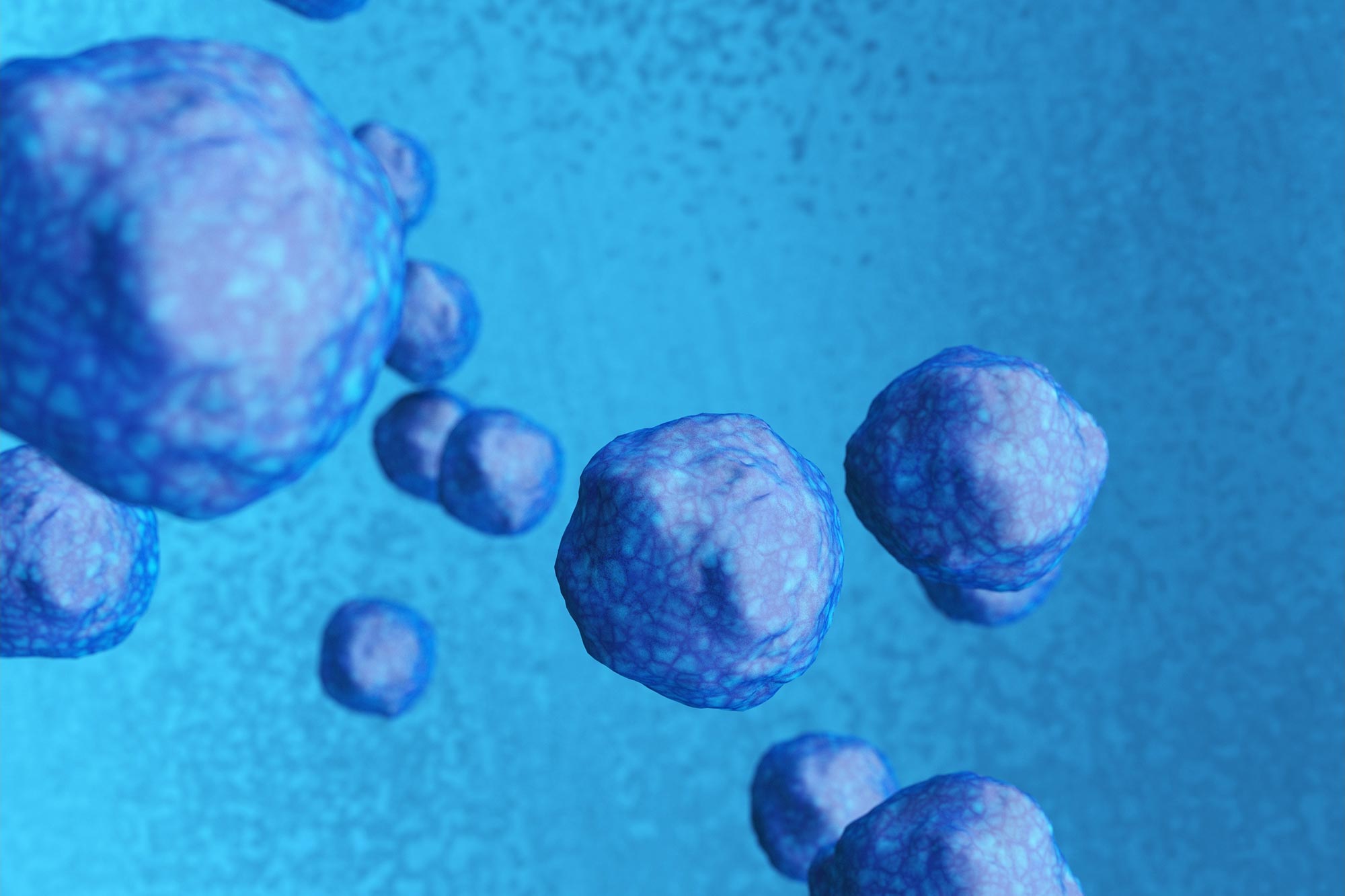
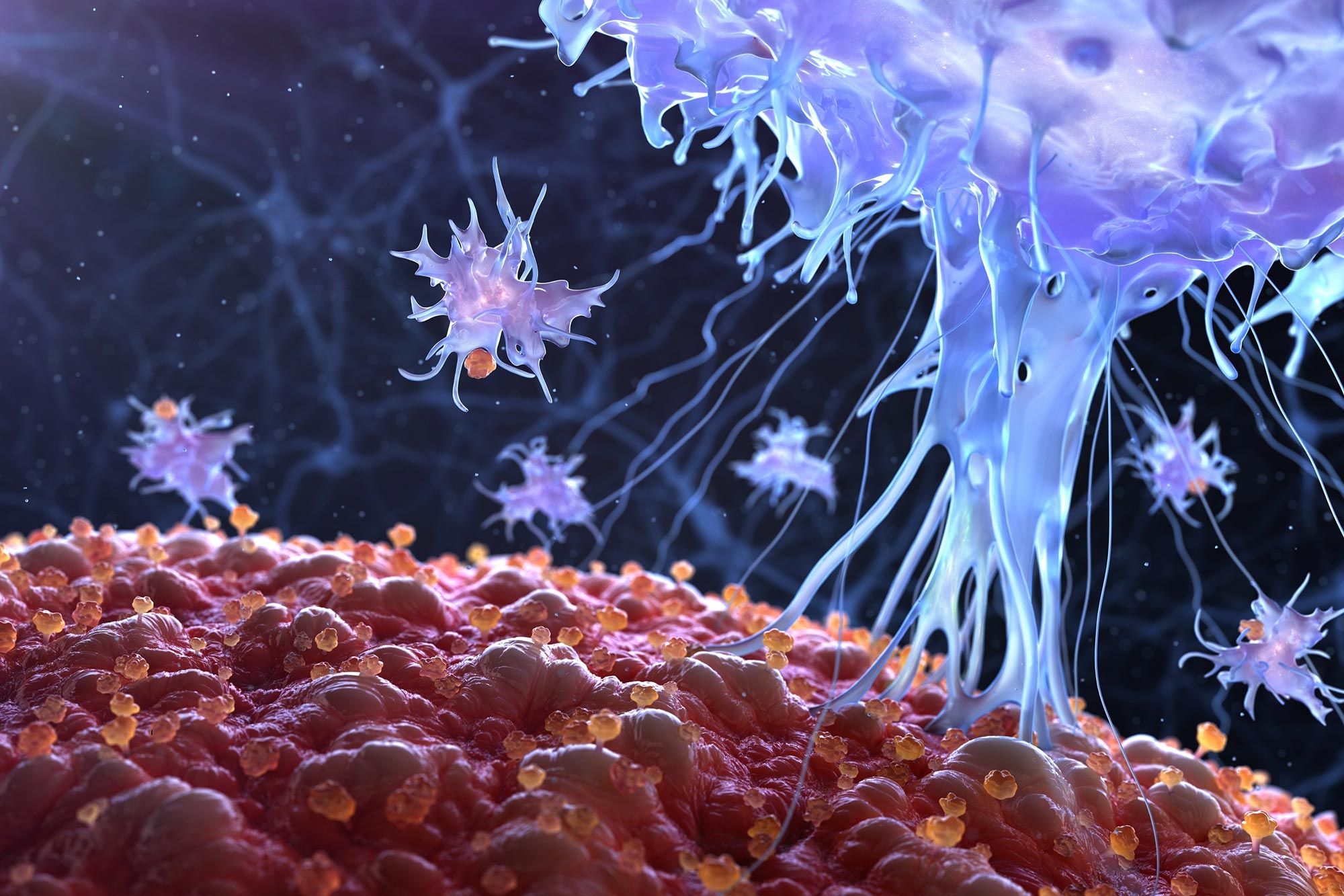
A team of scientists at Trinity College Dublin has discovered that a simple electric current can rewire one of the most important components of our immune system—the macrophage. By applying controlled electrical stimulation, these immune cells can be “persuaded” to suppress harmful inflammation and accelerate tissue repair. The work, published in Cell Reports Physical Science, signals the dawn of what could become an entirely new class of medicine: bioelectric healing.
Continue reading… “Using Electricity To “Reprogram” the Immune System for Faster Healing”